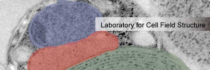
TOP > Research > Cell Dynamics Research Core > Laboratory for Cell Field Structure

Unit Leader
Atsuko Iwane (Ph.D.)
Electron microscopy (EM) offers the best in resolution of extremely small biological specimens, providing images of the minutest of organelles and molecules that are responsible for phenomena fundamental to life including cell division, differentiation and proliferation. The resulting fine structural information has been extraordinary in helping our understanding of how morphology and function relate.
We seek to image several target molecules and organelles, including the mitochondria and chloroplasts, and their surrounding environments inside a cell by using two different electron microscopy technologies. The first is Cryo-EM tomography, which produces a 3D image by scanning the field of view at multiple angles over an angular range from -70 degrees to 70 degrees relative to the perpendicular of the specimen plane used in transmission electron microscopy (TEM). The second is focused ion beam scanning electron microscopy (FIB-SEM), which uses SEM to scan a number of sections that are created by FIB. In this aim, we will combine our electron microscopy system with fluorescence microscopy to conduct dual imaging of the specimen in the same field of view. This will provide information on the dynamics of the specimen, which will complement the structure information and give us better understanding of the relationship between cell fate and cell morphology.
Research Topics
- Development of stainless cell imaging by Cryo-TEM
- Whole cell structure reconstruction by three-dimensional FIB-SEM
Selected Publications
- Fujii T, Iwane A.H., Yanagida T. Namba K.
Direct visualization of secondary structures of F-actin by electron cryomicroscopy
Nature, 467, 724-728, 2010 - Nishikawa S., Arimoto I., Ikezaki K.,Sugawa M., Ueno H., Komori T., Iwane A.H., Yanagida T
Switch between large hand-over-hand and small inchworm-like steps in myosin VI
Cell, 142, 879-888, 2010 - Watanabe T.M., Yanagida T., Iwane A.H.
Single molecular observation of self-regulated kinesin motility
Biochemistry, 49, 4654-4661, 2010 - Iwane A.H., Morimatsu M. Yanagida T.
Recombinant α-actin for specific fluorescent labeling
Proceedings of the Japan Academy, Series B, Physical and Biological Sciences, 85, 491-499, 2009 - Iwaki M., Iwane A.H., Shimokawa T., Coock R., Yanagida T.
Brownian search and catch mechanism for myosin-VI steps
Nature Chemical Biology, 5, 403-40, 2009 - Iwane A.H., Tanaka H., Morimoto S., Ishijima A., Yanagida T.
The neck domain of myosin II primarily regulates the actomyosin kinetics, not the stepsize
Journal of Molecular Biology, 353, 213-221, 2005 - Tanaka H., Homma K., Iwane A.H., Katayama E., Ikebe R., Yanagida T., Ikebe M.
The motor domain determines the large step of myosin-V
Nature, 415, 192-195, 2002 - Kitamura K., Tokunaga M., Iwane A. H., Yanagida T.
A single myosin head moves along an actin filament with regular steps of 5.3 nanometres
Nature, 397, 129-134, 1999 - Iwane A. H., Funatsu T., Harada Y., Tokunaga M., Yanagida T.
Single molecular assay of the individual ATP turnovers by a myosin-GFP fusion protein expressed in vitro
FEBS Letters, 407, 235-238, 1997 - Iwane A. H., Kitamura K., Tokunaga M., Yanagida T.
Myosin subfragment-1 is fully equipped with factors essential for motor function
Biochemical and Biophysical Research Communications, 230, 76-80, 1997