
EGFP and Protein G-coated Quantum dots enable in vitro/in vivo dual fluorescence imaging of tumors
May 1, 2015 Tweet
Recently, second-near infrared fluorescence (2nd NIR: 1000–1500 nm) has been used for non-invasive in vivo imaging. Second-NIR fluorescence imaging has advantages over traditional NIR fluorescence imaging (700–900 nm) such as reduced light scattering and minimal autofluorescence. Semiconductor quantum dots (QDs) such as PbS, PbSe, and Ag2S are promising fluorescent probes because of their strong fluorescence and photostability in the range of second-NIR.
However, fluorescence imaging using second-NIR QDs cannot achieve sub-cellular level resolution because of the long emission wavelength, and the fluorescence detectors of conventional microscope systems do not support second-NIR fluorescence imaging. Moreover, the preparation of the QD-based fluorescent probe needs multiple synthetic steps such as QD formation, surface modification and biomolecule conjugation, which sets a high hurdle for biologists. On the other hand conventional fluorescent proteins such as enhanced green fluorescent protein (EGFP) are widely used for sub-cellular level imaging using high resolution and high sensitivity microscopes but they are not suited for whole body imaging.
To compensate for the drawbacks of these probes, Akira Sasaki in QBiC Nano-Bioprobe research team (main affiliation: Biomedical Research Institute, National Institute of Advanced Industrial Science and Technology) has developed a dual (visible and second- NIR) emission QD probe that can be used for both in vitro cellular imaging and in vivo non-invasive whole-body imaging.
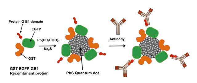
By coating QD with EGFP and protein G, which bind to immunoglobulin G, the team created an antibody-binding probe that emits visible green fluorescence and 2nd NIR fluorescence (1150nm). They administered the probe and anti-HER2 antibody, which recognizes a cell surface antigen increased in breast cancers, into nude mice that carry a breast cancer transplant, and succeeded in visualizing the cancer transplant non-invasively using the 2nd NIR fluorescence detector. They then excised cancer tissue and detected GFP signal on the cell surface by conventional confocal fluorescence microscopy.
Since the protein portions are produced as recombinant proteins by regular molecular biological techniques, researchers have the freedom to modify the protein portion according to their needs such as the color of the fluorescent protein. The paper also describes an easy one-step, one-pot procedure to synthesize the probe, thereby lowering the hurdle for biologists to use this new imaging methodology.
These results made the front cover of March 28 issue of Nanoscale.
- Sasaki A, Tsukasaki Y, Komatsuzaki A, Sakata T, Yasuda H, and Jin T. (2015) Recombinant protein (EGFP-Protein G)-coated PbS quantum dots for in vitro and in vivo dual fluorescence (visible and second-NIR) imaging of breast tumors.: Nanoscale, 7, 5115 doi: 10.1039/c4nr06480a
