
- 1
-
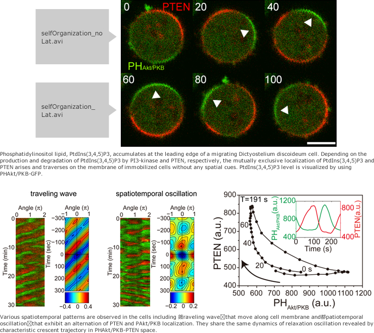
Spontaneous cellular polarity formation and self-organization
Self-organization in cellular polarity formation and migration
![]()
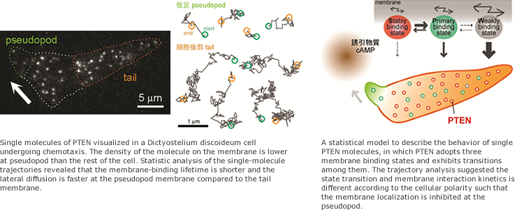
Single-molecule imaging of the components of the self-organization system
Individual PTEN molecules associate with the cell membrane only transiently (300 msec on average) at the region where a mass of the molecule exhibits localization. The quantification of the kinetics of membrane association/dissociation and PtdIns(3,4,5)P3 dephosphorylation is necessary for the understanding of the self-organized localization mechanism. We employ single-molecule imaging and tracking analysis combined with the techniques of molecular genetics and biochemical or structural biological experiments to clarify essential properties of stochastic molecular reactions and movements in the self organization.
![]()
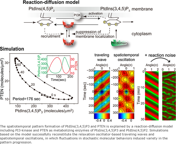
Interplay between experiments and a mathematical model
We have developed techniques of single-molecule imaging and simultaneous multicolor imaging in living cells and established methods of statistical analyses, elucidating stochastic behaviors of the individual molecules and averaged behaviors of the molecular ensemble. Quantitative data obtained from exact measurements can contribute toward understanding of an underlying mechanism with a help of mathematical models. Vigorous interactions among experiments, statistical analyses and model buildins are routine in our studies.
![]()
![]()
- 2
Sensing and signal tranduction
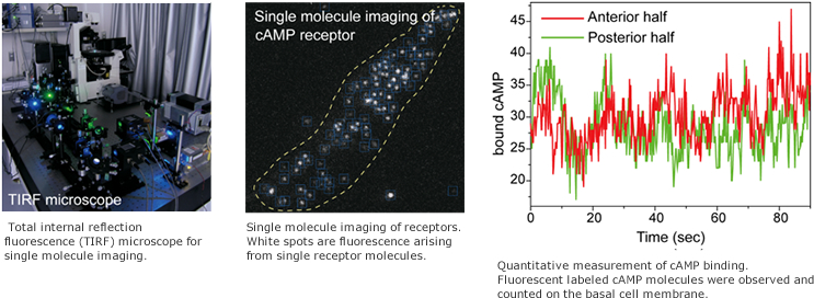
Single molecule imaging of a chemoattractant receptor.
Chemotactic signal transduction in Dictyostelium discoideum is mediated by a G protein coupled receptor. Cells sense the difference of the chemoattractant cAMP concentration between the front and back of the cell. In other words, they detect the difference in number of cAMP-bound receptor. Because the binding events between cAMP and the receptor are stochastic, the number of cAMP bound activated receptors has temporal dispersion and fluctuates around mean value. This mean value can be thought as a signal of the cAMP concentration, while the fluctuation is a detection error or noise in the cAMP signal. Cells can show proper chemotaxis in situation where noise exceeds the signal. How do cells achieve this? Single molecule imaging provides important clues to help answer that question. This technique allows visualization and quantification of signals and noises in cell signaling.
![]()
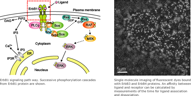
Regulation mechanism of cellular signaling by ErbB proteins
ErbB membrane receptors, which consist of four families designated ErbB1 - ErbB4, transmit extra-cellular signals into the cytoplasm. Because each ErbB protein interacts with family-specific ligands and activates a receptor-specific signaling pathway, a cell reaction depends on the extracellular ligand-receptor combination. For example, ErbB1 which binds the epidermal growth factor (EGF) ligand is also called the EGF receptor (EGFR) and a principal receptor controlling cell proliferation. If the regulation of ErbB1 expression or receptor function is lost, extraordinary proliferation is induced that leads to serious diseases such as cancer.
Single-molecule imaging of ErbB proteins conjugated with a fluorescent probe enables a direct observation of the individual receptors on the plasma membrane. By analyzing motility or brightness of fluorescent spots in the acquired images, we obtain detailed information about ErbB including its molecular behaviors, interactions with ligands, and reactions with other signaling proteins.
Elucidating the regulatory processes in cellular signaling based on this information, we aim to understand the mechanism of cellular reactions and initiation / progression of diseases.
![]()
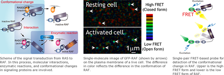
Single-molecule measurement of intracellular signaling from RAS to RAF and cell-fate determination
Uncontrollability of cellular behaviors is a major obstacle to the therapeutic application of regenerative biology. To regulate cellular behaviors, it is important to understand how cellular system work and to find the key factors to monitor and manipulate the states of the cellular system. Here, we focus on the signaling molecule RAS and RAF as one of the candidates of the key factors to monitor and predict cellular behaviors. RAS-RAF signaling regulates various cellular responses including proliferation, differentiation, and carcinogenesis. By using single-molecule imaging techniques, we study the spatiotemporal dynamics of the movement, the interaction of RAS and RAF, the enzymatic reaction, and the conformational changes in RAF in living cells.
![]()
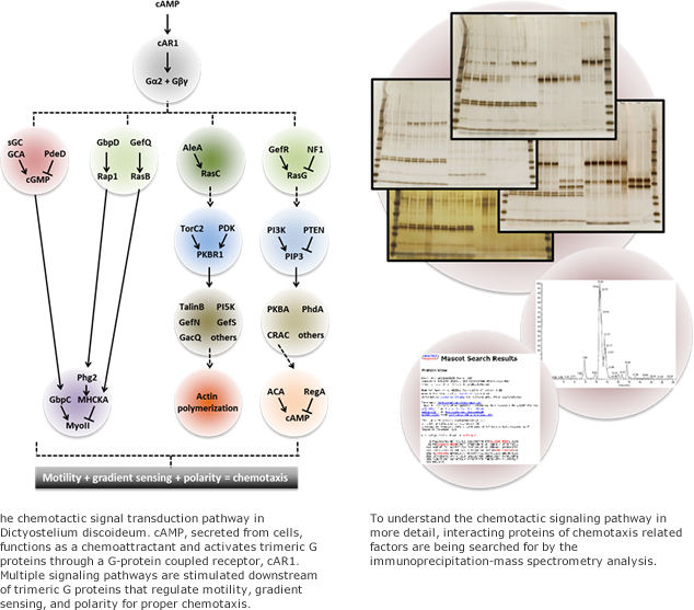
Signaling network of chemotaxis
Cells can direct their movement according to external chemical gradients. This directional motility is known as chemotaxis and observed in many biological situations including embryogenesis, neuronal pattern formation, and metastasis of cancer cells. To understand the mechanisms of how cells process signals of chemical gradients to control their directional motility, we are striving to reveal the signaling network in chemotaxis and the reaction dynamics with quantitative data of each composing module. These studies could be expected to give a new insight into flexible biological systems and result in applicable technologies for engineering and medical fields.
![]()
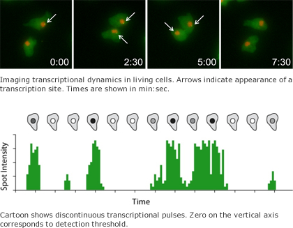
Noisy transcription
Recent progress in imaging transcription in individual living cells has revealed that gene transcription occurs in pulses or bursts. We would like to understand the biological origins of transcriptional bursting and how it is regulated by chromatin structure. We are also trying to understand how noisy transcription is related to differentiation in development.
![]()
![]()
- 3
Quantitative measurement technology
Cell tracking in a multicellular tissue
Cells aggregates organize themselves into functional tissues during metazoan development. By analyzing the collective cell migration of Dictyostelium aggregates and the processes by which they are transformed into highly structured tissues such as the stalk and fruiting body, we attempt to elucidate the mechanisms of systematic tissue formation. We believe that these studies will allow us to understand conserved principles underlying embryo morphogenesis and regeneration processes in metazoans.
![]()

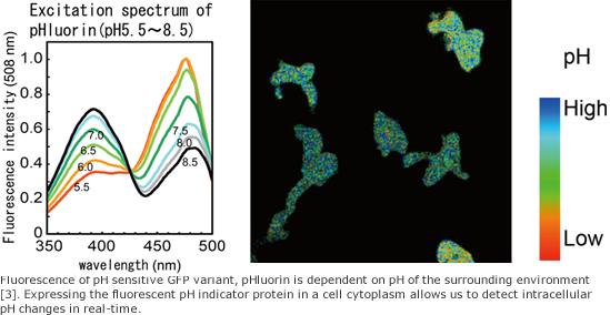
Intracellular pH imaging
Intracellular pH plays an important role in signal transduction. For example, it has been reported that the migration speed of cells is highly correlated with intracellular pH [1,2]. To investigate the role of intracellular pH in signal transduction, we image and measure the intracellular pH of living cells. Additionally, it is also known that cancer formation is associated with intracellular pH changes. A greater understanding the role of pH in signal transduction may contribute to new medical technologies.
References- [1]
- Simchowitz L. & Cragoe E. J. Jr. J. Biol. Chem. 261‚ 6492-6500. (1986)
- [2]
- Van Duijn B. & Inouye‚ K. PNAS 88‚ 4951?4955 (1991).
- [3]
- Miesenbock G‚ et al.‚ Nature 394‚ 192?195‚ (1998).
![]()
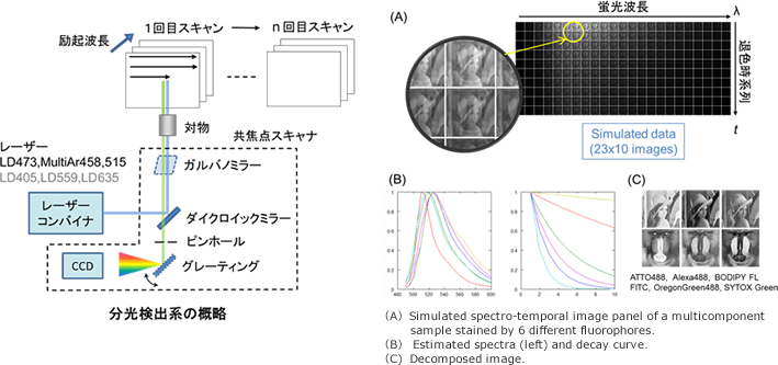
Polychromatic fluorescence imaging
It is valuable to detect multiple fluorescent probes simultaneously in order to analyze co-varying intracellular components. The technique of spectroscopic imaging combined with linear unmixing, in principle, allows more than 10 colors to be simultaneously imaged. However, there are factors limiting the widespread use of spectroscopic imaging which include the requirement for singly stained reference samples, and difficulty in accurately separating auto-fluorescent signals. We are developing a new polychromatic imaging technique which overcomes these limitations.
![]()